Wednesday, September 23, 2009

Split notochord syndrome

Dr Halim wrote:

Split notochord syndrome

Split notochord syndrome(SNS)

Split notochord syndrome is a spectrum of congenital spinal malformations that develops due to an adhesion between endoderm and ectoderm causing the "splitting" of notochord due to a persistent connection between endoderm and dorsal ectoderm.

  1. Dorsal enteric fistula,
  2. dorsal sinus,
  3. dorsal diverticula and
  4. neurenteric cysts are the components of the spectrum
  5. Diastematomyelia

The SNS, as proposed by Bentley and Smith, (also known as posterior spina bifida, combined spina bifida, neurenteric fistula, dorsal enteric fistula) is an extremely rare form of dysraphism It was first described by Rembe in 1887. In this syndrome, vertebral anomalies (anterior and posterior spina bifida, butterfly vertebrae), central nervous system abnormalities (diastematomyelia, diplomyelia, myelomeningocele) and intestinal anomalies (fistulas, dermal sinus tract, diverticula and enteric cysts) are associated with each other.

The syndrome manifests as a cleft in the dorsal midline of the body through which intestinal segments are exteriorized (often with an associated fistula), myelomeningocele, and occasionally as a teratoma. Central nervous system abnormalities are always present: hydrocephalus and diastematomyelia/diplomyelia are constant findings. However, babies do not necessarily present with functional spinal cord defects: in some reported cases, the motor function of the lower limbs is normal. Talipes equinovarus is a common finding.

Diastematomyelia is a form of split notochord syndrome with sagittal division of the spinal cord into two pia-lined hemicords. In complete clefts with formation of two dural sacs, a fibrous, cartilaginous or osseous spur is present in half of the cases. The doubling of the cord can occur in the Thoracic region

All forms of split notochord syndrome are frequently associated with vertebral anomalies such as anterior and posterior spina bifida, butterfly vertebrae and hemivertebrae

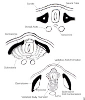
The embryologic mechanism for the development of split notochord syndrome was first discribed by Saunders in 1943 In the pathogenesis of split notochord syndrome, an adhesion occurs between endoderm and ectoderm in the route of notochord and notochord splits around the adhesion creating a defect in the vertebral column. Adhesion between endoderm and ectoderm also results in a potential connection between yolk sac (gut) and dorsal ectodermal surface (skin). This connection persists as a tractus in the dorsal enteric fistula, which is the most severe form of split notochord syndrome or may obliterate at any point creating a dorsal enteric sinus, diverticula and neurenteric cysts

The location of neurenteric cysts may be pre-vertebral, intraspinal or post-vertebral. The posterior mediastinum is a frequent location for pre-vertebral neurenteric cysts. Neurenteric cysts are lined by gastrointestinal or respiratory epithelium. Anterior and posterior spina bifida, butterfly vertebrae and hemivertebrae frequently accompany the neurenteric cysts . Prevertebral neurenteric cysts may be connected to the meninges or spinal cord by a tube or a fibrous neurenteric band via an anterior spina bifida and dura defect

NB
Picture taken from - www.scielo.br/.../jped/v80n1/en_80n1a15f01.gif


Spine Embryology

Development of the spine

Molecular and cellular tissue interactions, as well as increasing organ complexity, characterize the fundamental features of the embryonic developmental process during axial embryogenesis. Alteration in the molecular and macromolecular process may lead to structural defects involving the spine and spinal cord. Such defects may occur prenatally and/or postnatally and, from the standpoint of basic developmental pathogenesis, can be divided into the following 3 categories:

  • Malformation
  • Disruption
  • Deformation

Malformation

Malformation is a failure of the embryologic differentiation and/or development of a specific anatomic structure, causing it to be absent or improperly formed before the fetal period commences. (An example is the formation of a hemivertebra.) Once it is established anatomically, the defect may continue to affect spinal development adversely throughout the subsequent fetal and postnatal periods. The type of malformation and its severity depend on the stage of the developmental or maturation cycle that is specifically affected.

Disruption

Disruption refers to the destruction of an anatomic feature that formed normally during the embryonic period. This phenomenon, resulting in a structural defect, involves the limbs (amniotic band syndrome) more frequently than the spine, during the fetal stage.

Deformation

Deformation is an alteration in the shape or structure of an individual vertebra or of the entire spine during the fetal and/or postnatal periods, after the involved region's initial, normal differentiation. Infantile scoliosis and adolescent scoliosis are examples of conditions resulting from deformation and are therefore not true congenital forms of scoliosis. Deformations may be divided into those that are intrinsically derived and those that are extrinsically derived.

Intrinsic deformations result from the reduced ability of the fetus or child to move away from normal imposed forces and depend on the integrity of the neuromuscular system to respond effectively. Extrinsic prenatal deformations are the result of reduction in the amount of space in which a developing fetus may move. Such reduction may be either physiologic or pathologic.

Physiologic structural deformation usually is limited and relatively reversible once the constraining influences are removed. In contrast, pathologic structural deformation is a more permanent process.

Late gestational deformations have an excellent prognosis. Approximately 90% of such deformations noted at birth correct spontaneously. The earlier a deformation is recognized, the greater the likelihood of correcting it or at least preventing further deformation (with, for instance, early bracing for idiopathic scoliosis).

Embryogenesis

Gestation has been divided into the embryonic period and the fetal period. The embryonic period is considered to be the time from fertilization to the end of the eighth week of gestation. The remainder of gestation is called the fetal period. By the end of the embryonic period, all of the major organ systems have been established, and the basic body plan is complete. The 5 major stages of embryonic development can be summarized as follows:

  • Fertilization - Female and male gametes combine to form a zygote.
  • Cleavage - The zygote divides into a ball of smaller cells, each receiving different parts of the maternal cytoplasm.
  • Gastrulation - The cells migrate and proliferate to form the 3 primary germ layers, namely, the ectoderm, the mesoderm, and the endoderm.
  • Neurulation - The notochord, neural crest, and precursors of the central and peripheral nervous system form.
  • Organogenesis - Primary cell types differentiate to generate the organs.


The spine has its embryologic origins in the cell that is induced to migrate out of the somite and toward the notochord and the neural tube . As a mass of sclerotomal cells collects segmentally at the embryonic midline, surrounding the neural tube and the notochord, the sclerodermal cells begin to separate into a cranial portion and a caudal portion.

The cranial portion of each sclerotome recombines with the caudal portion of the directly superior sclerotome in a resegmentation process known as metameric shift.After the metameric shift, spinal nerves, which originally left the neural tube to go to the center of the sclerotome, are able to pass between the precartilaginous vertebral bodies to innervate the segmentation myotomes

The atlas and axis form by a mechanism that is different from that of the other vertebral bodies. Part of the first cervical sclerotome plus the cranial portion of the second cervical sclerotome contribute cells, forming the odontoid process and the arch of the atlas.

In the cervical region, the 8 cervical somites generate 7 cervical vertebrae because the cranial portion of the first cervical sclerotome contributes to the formation of the occiput and the caudal portion of the eighth cervical sclerotome contributes to T1. In this way, the 8 cervical spinal nerves become associated with 7 cervical vertebrae The first cervical spinal nerve passes between the base of the skull and the first cervical vertebra. The eighth cervical nerve exits below the seventh cervical vertebra and above the first thoracic vertebra. The remainder of the nerve roots exit below their corresponding vertebral bodies.

The intervertebral disks form in the area between the resegmented vertebral bodies after the split of the sclerotomes. The nucleus pulposus originates from cells of the notochord, whereas the annulus fibrosis originates from the sclerotomal cell.

At the same time that the sclerotomes are undergoing a shift to form the vertebral bodies, the heart, kidneys, trachea, and esophagus are differentiating, which means that a noxious influence during this time can affect the adjacent, simultaneously developing organs. Hence, cardiac anomalies often are associated with congenital scoliosis of the thoracic spine, and renal anomalies are often associated with congenital malformation of the lumbar spine. VACTERL syndrome (abnormalities of the vertebrae, anus, cardiovascular tree, trachea, esophagus, renal system, and limb buds) is an extreme example of association of malformation in the embryonic stage.

Genetics

A family history of congenital spinal deformity is rare. Winter and colleagues found that only 13 of 1250 patients had a positive family history of such deformity Studies of twins usually have shown that if 1 twin has an anomaly, the other does not, even if the twins are identical. However, several investigators have described hereditary congenital scoliosis. Most of the patients in those reports had extensive defects of segmentation in association with spondylocostal, costovertebral, or spondylothoracic dysplasia (Jarcho-Levin syndrome). In addition to spinal segmentation defects, segmentation defects often occur in the ribs, leading to the development of a small, stiff thorax and, frequently, to pulmonary compromise. Such defects also may extend into the cervical spine as a Klippel-Feil anomaly.

Article taken from

medscape

Congenital Spinal Deformity

Author: Robert Mervyn Letts, MD, FRCS(C), FACS, Former Chief, Department of Surgery, Division of Pediatric Orthopedics, Children's Hospital of Eastern Ontario, University of Ottawa; Consultant Pediatric Orthopedic Surgeon, Sheikh Khalifa Medical City, UAE

Coauthor(s): Ayman Hussein Jawadi, MBBS, Pediatric Orthopedic Consultant, Department of Surgery, King Fahad National Guard Hospital

Atlanto axial instability in Down syndrome


(Our patient presented with torticollis)

Atlantoaxial instability (AAI), which denotes increased mobility of C2 in relation to C1, occurs more frequently in persons with Down syndrome (DS) than in the general population. The association was first reported in 1961 almost 100 years after DS was described. AAI, which affects 10–20% of individuals with DS, is mostly asymptomatic and is diagnosed using radiography

The instability is recognized through lateral neck radiographs by the presence of an abnormally large anterior atlanto-odontoid distance (AAOD)
The upper limit of a normal AAOD is 4 mm in subjects who are younger than 15 years and 3 mm in older subjects The anterior space between the atlas and the axis normally opens in flexion and closes in extension, which is why the AAOD is greater when the neck is radiographed in flexion than it is in neutral or extension positions The danger of AAI is that it becomes symptomatic in 1– 2% of individuals with DS when the displaced odontoid impinges the spinal cord .
Manifestations include

  • neck discomfort,
  • abnormal gait,
  • change in sphincteric control,
  • upper motor neuron lesion (UMNL),
  • paralysis, and even death.
In over 80% of symptomatic patients, this is the end result of chronic gradual instability In the remaining patients, where it may be preceded by a normal radiograph, it can be precipitated by neck trauma, sports injury, endotracheal intubation, or head and neck surgery

Patients with symptomatic AAI need urgent evaluation and management, which may include posterior fusion of C1 to C2. Patients with the asymptomatic condition need to take precautions to avoid neck injury as well as regular followups to detect any neurological deterioration.

The occurrence of degenerative changes in the cervical spine of patients with DS was first reported in 1966 As expected, these changes increase with age but they occur earlier than in the normal population Although cervical spondylosis has the potential to cause cervical cord damage, it has received less attention in the literature than AAI. This is mainly because most of the studies about cervical spine abnormalities were based on paediatric populations.

Abnormalities of the odontoid peg, such as
  • odontoid hypoplasia and
  • the presence of accessory ossicles
  • Os odontoideum may contribute to AAI.
Odontoid hypoplasia may cause slippage of the transverse ligament over the superior aspect of the shortened odontoid . Os odontoideum, which appears radiographically as a radiolucent oval ossicle with a smooth dense border of bone, causes loss of strength of the odontoid process as a post for attachment of ligaments .
AAI had degenerative changes in the cervical spine may implicate ligamentous laxity as a common mechanism. Lax ligaments allow more than normal motion between vertebral bodies and may lead to early disc lesions. Identification of cervical spondylosis in DS patients with AAI is important. More interesting than a common aetiology between the two conditions is the potential functional interaction. The restriction of motion lower in the cervical spine, brought on by degenerative changes, places stress on the extremely
mobile atlantoaxial joint and may increase the instability. If the prevalence of AAI decreases with advancing age, more attention should be paid to cervical spondylosis, which increases with age, in adults with DS.
The American Academy of Pediatrics has recommended that all children with Down syndrome get screening neck x-rays between the ages of 3 and 5 years, but this recommendation has not been universally embraced. It does seem prudent to obtain x-rays in affected children prior to general anesthesia (where the neck is bent backward prior to intubation), or if the child is considering participation in contact sports.

Other spine problems with Down syndrome:
Scoliosis

Issues in management of Down Syndrome
  • Mental retarded hence the ability to follow instructions and the frequency of self-inflicted trauma
  • Getting a proper implant sizes(small vertebrae) hence the need to use jacket such as minerva jacket
  • The presence of other congenital anomalies such as VSD/ASD or congenital spines which complicates the management or other skeletal problems such as hip dislocation
References
Cervical spine abnormalities associated with Down syndrome
Fawzi Elhami Ali et alInternational Orthopaedics (SICOT) (2006) 30: 284–289
DOI 10.1007/s00264-005-0070-y

Thursday, September 10, 2009

Process Uncinatus

It is not a true joint.It is a developmental bone projection from posterior lateral aspect of superior portion of the vertebral bodies,do not appear until the latter part of the first decade.


In general animal that hold their necks in an upright position have this process.There is a small gap between the uncus and the opposite surface of the upper spine.It is called lateral joint by Luschka. But there is no disc formed because uncus belongs to the arch and not to the body and the disc


can only be formed in correspondence to the body
When viewed anteriorly the lateral margin of the superior surface of each subaxial cervical vertebral body extends cranially as a bony process called the uncinate process.It articulates with a reciprocal convex area on the inferolateral aspect of the next cranial vertebral body.
This articulation is called uncovertebral joint or neurocentral joint of Luschka

It serves as
  • rail to limit lateral translation or bending and as a guiding mechanism for flexion and extension.
  • as a barrier to the direct extrusion of the disc material posterolaterally into the foramen and against the nerve root and vertebral artery
Surgically it is important because:
  • It is involves in degenerative changes and formation of osteophytes .It can compressed on the vertebral artery when it projects into the interverbral foramen and the artery is which already fixed in the vertebral foramen
  • It is a landmark(its medial border) during corpectomy to avoid injuring the vertebral artery and a landmark before operating microscope is positioned.
  • It anterior decompressive surgery for OPLL,it is important to exposed widely both uncus to confirm the localization of OPLL to the body
  • Pseudofracture of uncinate process is a known radiological phenomenon due to Mach effect

References:
Clinical anatomy of cervical spine: k hayashi -chapter 2 in book"Cervical spondylosis and similar disorders"
Cervical spine 3rd Edition CSRS